
Application of argon ion polishing-scanning electron microscope in the study of concrete microscopic pore structure
|
WANG Tao,male,born in 1992,engineer,focusing on microbeam analysis research such as scanning electron microscopy. E-mail:15901029788@163.com |
Received date: 2025-01-01
Revised date: 2025-01-18
Online published: 2025-11-07
Supported by
National Innovation Platform Open Fund Project of China Academy of Railway Sciences Corporation Limited(2022YJ119)
(2022YJ119)
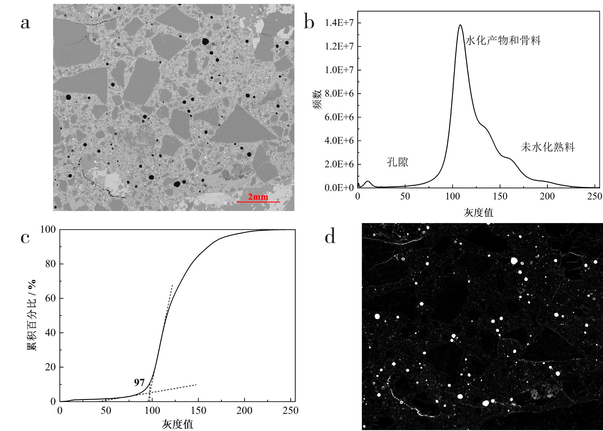
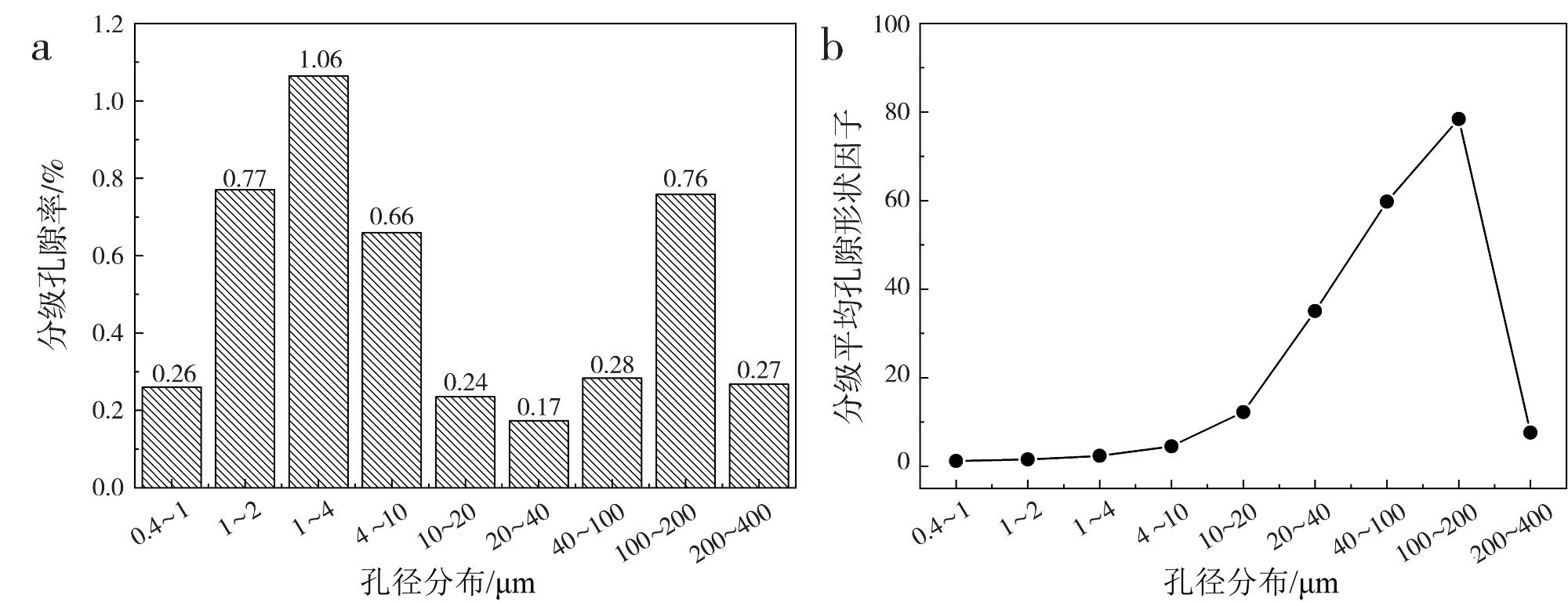
The microscopic pore structure is of great significance to the study of concrete materials. However,the traditional pore structure testing methods, such as nuclear magnetic resonance (NMR),mercury intrusion porosimetry (MIP),optical microscopy,X-ray tomography (X-CT),and nitrogen adsorption,still have some limitations in terms of pore morphology and nanoscale pore characterization. In this study,we proposed to use argon ion polishing-scanning electron microscopy method to study the microscopic pore structure in concrete,prepared concrete samples with high-quality mechanically damage-free surfaces, and qualitatively investigated four types of pore structure in concrete. Large-area backscattered electron images were collected, and the pore porosity,pore diameter,pore shape factor,and graded porosity of the pore structure were quantified by ImageJ,Avizo,and other software. This method achieves the qualitative analysis and quantitative characterization of concrete microporous structure, which is of reference significance for the research and analysis of the microstructure of concrete materials.
Tao WANG , Shengli CHEN , Xiangkun GE . Application of argon ion polishing-scanning electron microscope in the study of concrete microscopic pore structure[J]. World Nuclear Geoscience, 2025 , 42(1) : 196 -202 . DOI: 10.3969/j.issn.1672-0636.2025.01.017
表1 混凝土配合比/(kg·m-3)Table 1 Mix proportion of concrete/(kg·m-3) |
| 编号 | 水泥 | 粉煤灰 | 细骨料 | 碎石(5~10 mm) | 碎石(10~20 mm) | 减水剂 | 水 | 引气剂 |
|---|---|---|---|---|---|---|---|---|
| BY | 400 | 80 | 723 | 770 | 333 | 5.74 | 134 | 4.78 |
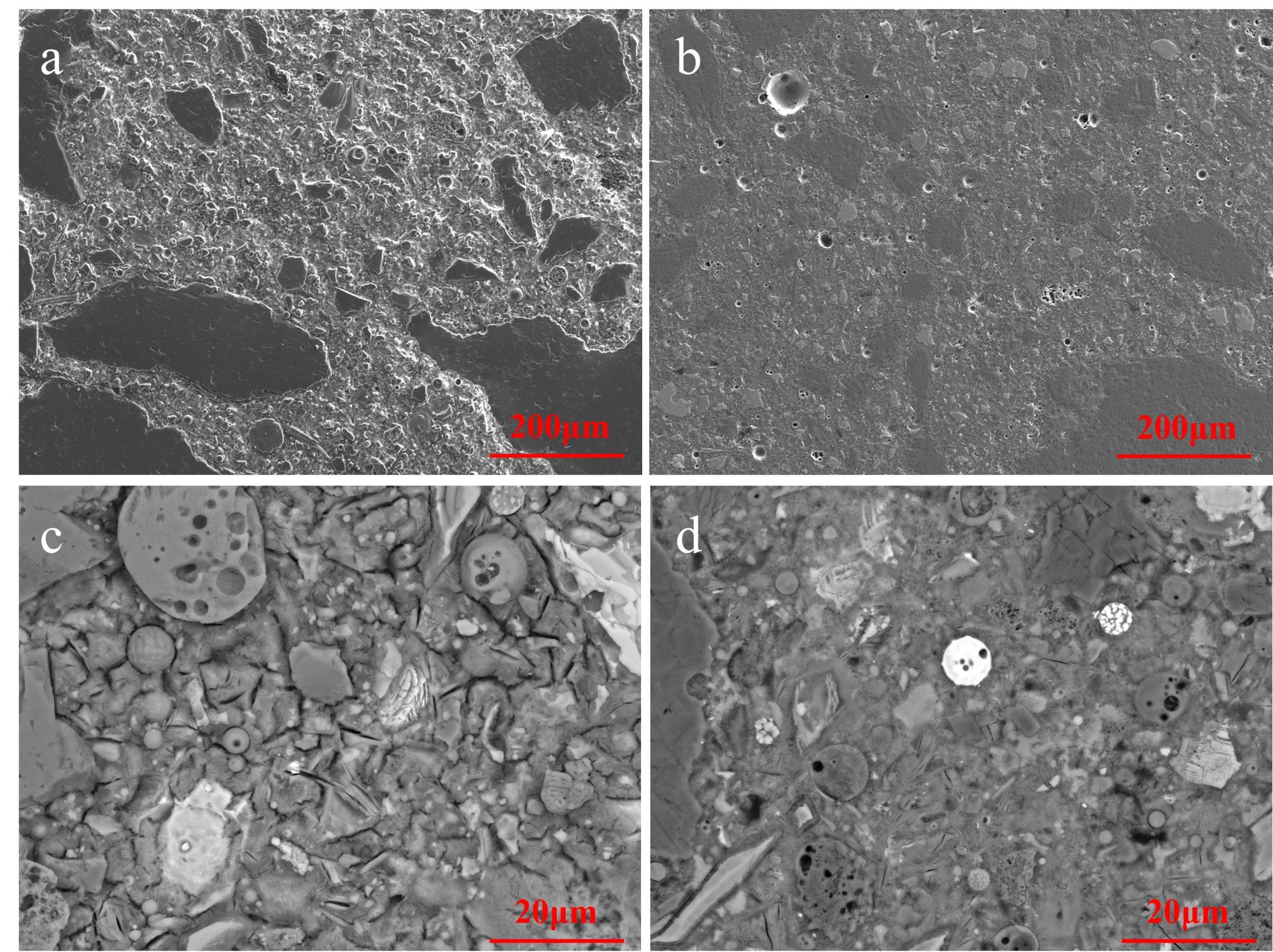
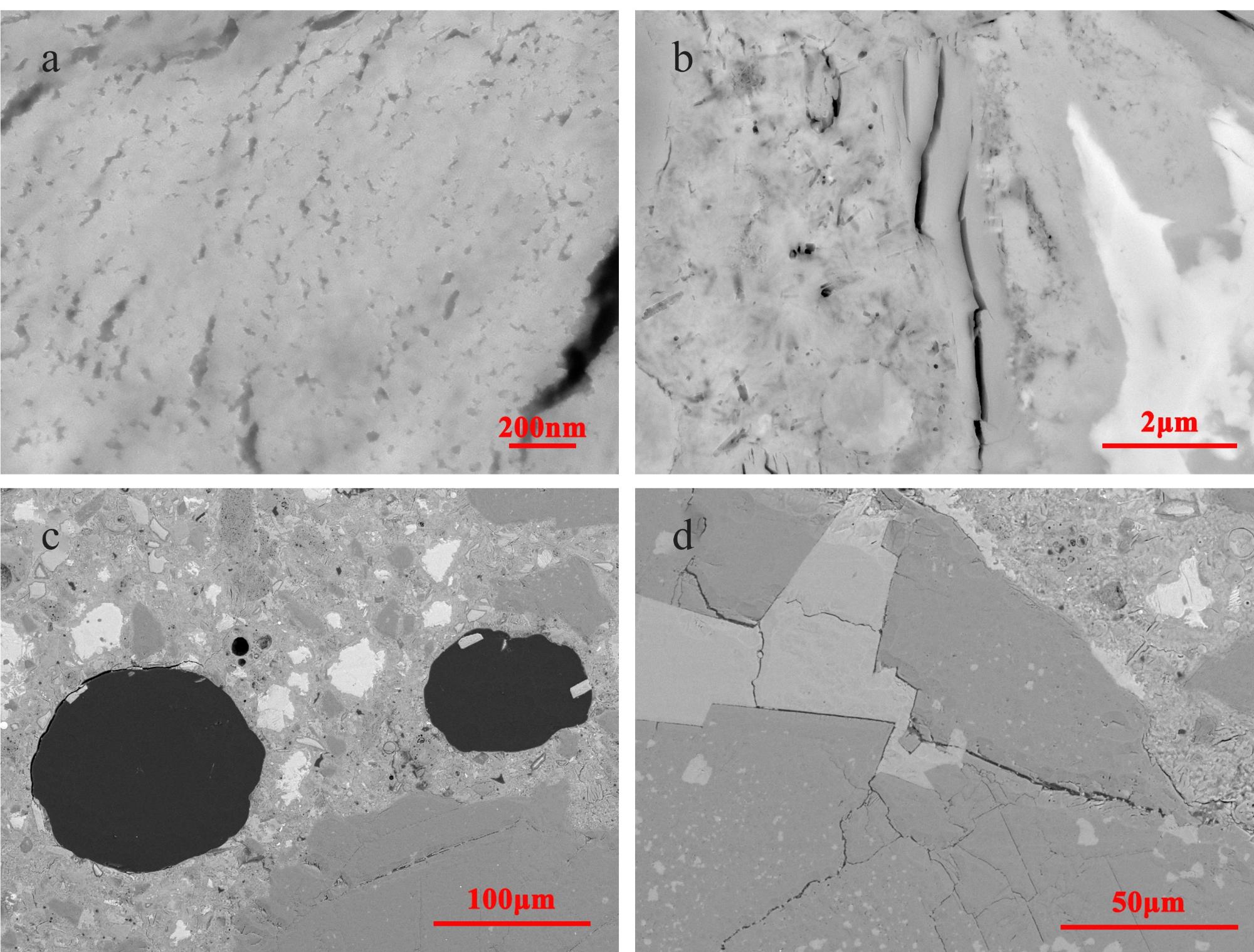
图1 不同抛光方式后样品扫描电镜图像a—机械抛光后SE图像;b—氩离子抛光后SE图像;c—机械抛光后BSE图像;d— 氩离子抛光后BSE图像。 Fig. 1 Scanning electron microscope images of samples after different polishing methods a-SE image after mechanical polishing;b-SE image after argon ion polishing;c-BSE image after mechanical polishing;d-BSE image after argon ion polishing. |
表2 混凝土孔隙结构参数Table 2 Pore structure parameters of concrete |
| 编号 | 孔隙直径/μm | 平均孔隙直径/μm | 孔隙面积/μm2 | 孔隙率/% | 平均孔隙形状因子 |
|---|---|---|---|---|---|
| BY | 0.45~300 | 1.75 | 4 794 940.62 | 4.47 | 1.71 |
| 1 |
陈茹梦. 人造骨料特性对混凝土界面过渡区的影响研究[D]. 长沙: 湖南大学, 2021.
|
| 2 |
胡曙光, 袁盼, 王发洲, 等. 背散射电子图像分析法在水泥基材料孔结构研究中的应用[J]. 建筑材料学报, 2017, 20(2):316-320.
|
| 3 |
|
| 4 |
|
| 5 |
高辉, 张雄, 张永娟. 混凝土气孔结构对其强度及界面过渡区的影响[J]. 同济大学学报(自然科学版), 2014, 42(5):751-756.
|
| 6 |
高翔. 水泥基材料微观表征技术的研究及应用[D]. 南京: 东南大学, 2018.
|
| 7 |
陈杰, 周改英, 赵喜亮, 等. 储层岩石孔隙结构特征研究方法综述[J]. 特种油气藏, 2005, 12(4):11-5.
|
| 8 |
陈一呜, 魏秀丽, 徐欢. 北美页岩气储层孔隙类型研究的启示[J]. 复杂油气藏, 2012, 5(2):19-22.
|
| 9 |
|
| 10 |
王羽, 金婵, 汪丽华, 等. 应用氩离子抛光-扫描电镜方法研究四川九老洞组页岩微观孔隙特征[J]. 岩矿测试, 2015, 34(3):278-285.
|
| 11 |
|
| 12 |
|
| 13 |
邓刘敏, 葛祥坤, 范光, 等. 基于扫描电镜-能谱仪的矿物定量分析——以 AMICS 为例[J]. 世界核地质科学, 2023, 40(1):98-105.
|
| 14 |
|
| 15 |
周晋辉, 郭晓阳, 赵博, 等. 煤岩体系微观组分及孔隙结构对甲烷吸附的影响研究[J]. 矿业安全与环保, 2024, 51(1): 51-60.
|
| 16 |
|
/
| 〈 |
|
〉 |